
Identification of Librela Associated Adverse Joint Events
 Click to view full screenAndrew Armitage BSc BVM&S MRCVS
Affiliations
Greenside Referrals
Regenerative Medicine and Rehabilitation
St Boswells, Scotland
Please Click here to view the full publication
Please click here to view the research poster
Introduction
Librela (bedinvetmab), a monoclonal antibody for managing osteoarthritis pain in dogs, generally has a favourable safety profile reported. Recently, the FDA’s Center for Veterinary Medicine evaluated adverse events reported in dogs treated with Librela. These events included ataxia, seizures, neurologic signs, paresis, recumbency, urinary incontinence, polyuria, and polydipsia. In some cases, death or euthanasia was reported as an outcome.
Adverse events concerning the musculoskeletal system in dogs are underreported. However, the FDA put human anti-NGF mAb development programs on hold due to increased serious joint-related adverse events. These included osteonecrosis and rapid progressive osteoarthritis, occurring in patients treated with tanezumab alone or with NSAIDs. These events were noted in phase II and III trials, with a mean treatment duration of 199 days (1). Higher incidence of joint destruction was linked to longer exposure, larger doses, and concurrent NSAID use (2,3), though serious joint issues also occurred in some after a single anti-NGF mAb treatment (4).
Characteristics of RPOA are rapid clinical deterioration (increase in pain) and radiographic progression of joint degeneration (5). Although theories have been proposed, the cause of anti-NGF related RPOA remains unclear (8). Overloading, resulting from increased activity and weight-bearing due to good analgesia (analgesic arthropathy), immune reactions and neuropathic arthropathy (nerve damage resulting in loss of ability to feel the joint and decreases joint stability) have been suggested as potential factors leading to RPOA following anti-NGF therapy (4,9,10).
There are now reports of similar joint adverse events (AE) occurring in dogs receiving long term Bedinvetmab treatment but the pathology has yet to be defined.
Please Click here to view the full publication
Please click here to view the research poster
Objective
To define and identify the appearance of atypical osteoarthritis and periarticular new bone formation in elbow joints of canine patients receiving long term Librela administration.
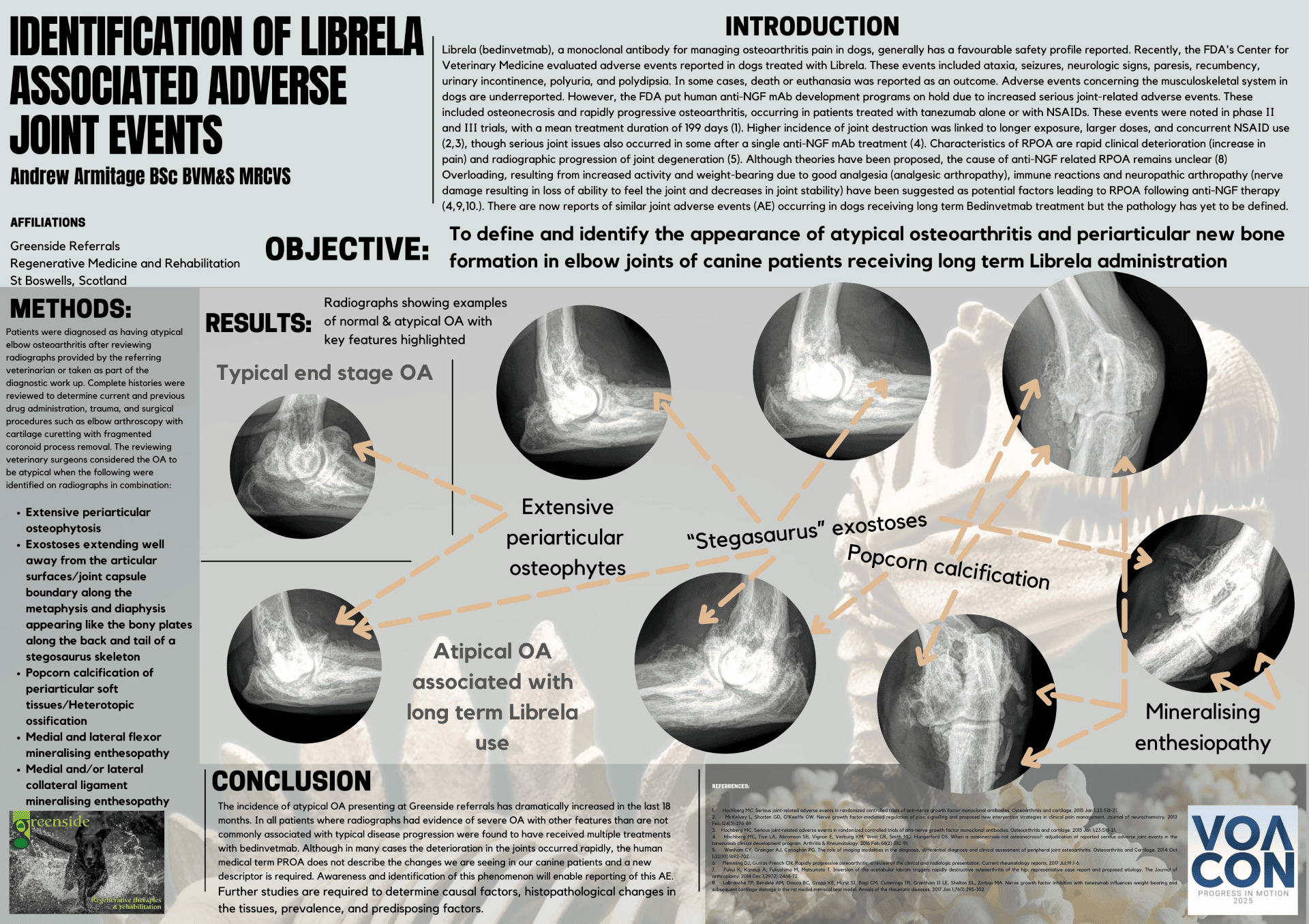
Methods
Patients were diagnosed as having atypical elbow osteoarthritis after reviewing radiographs provided by the referring veterinary surgeon or taken as part of the diagnostic work up. Complete histories were reviewed to determine current and previous drug administration, trauma, and surgical procedures such as elbow arthroscopy with cartilage curetting with fragmented coronoid process removal. The reviewing veterinary surgeons considered the OA to be atypical when the following were identified on radiographs in combination:
- Extensive periarticular osteophytosis
- Exostoses extending well away from the articular surfaces/joint capsule boundary along the metaphysis and diaphysis appearing like the bony plates along the back and tail of a stegosaurus skeleton
- Popcorn calcification of periarticular soft tissues/heterotopic ossification
- Medial and lateral flexor mineralising enthesopathy
- Medial and/or lateral collateral ligament mineralising enthesopathy
Results
Radiographs showing examples of normal & atypical OA with key features highlighted,
Typical end stage OA
Extensive periarticular osteophytes
“Stegosaurus” exostoses
Popcorn calcification
Atypical OA associated with long term Librela use
Mineralising enthesiopathy
Conclusion
The incidence of atypical OA presenting at Greenside referrals has dramatically increased in the last 18 months. In all patients where radiographs had evidence of severe OA with other features than are not commonly associated with typical disease progression were found to have received multiple treatments with bedinvetmab. Although in many cases the deterioration in joints occurred rapidly, the human medical term RPOA does not describe the changes we are seeing in our canine patients and a new descriptor is required. Awareness and identification of this phenomenon will enable reporting of this AE. Further studies are required to determine causal factors, histopathological changes in the tissues, prevalence, and predisposing factors.
Please Click here to view the full publication
Please click here to view the research poster