
Dirsko J.F. von Pfeil 1,2,3 | Andrew Armitage 4 | Nathan C. Nelson 5
Bedinvetmab and frunevetmab are monoclonal antibodies targeting nerve growth factor (aNGFmab), designed to alleviate pain associated with canine or feline osteoarthritis. They inhibit nerve growth factor (NGF)-mediated nociception and peripheral sensitization. They have been approved in multiple countries since 2020, and several studies have shown good responses. These medications have been used successfully in millions of pets worldwide.
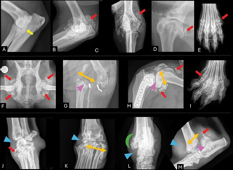
Fig. 1. Radiographic appearance of various joints of dogs that received bedinvetmab (Librela®/Berensa®). Elbows: Diagnosis of medial compartment syndrome and radiographic changes with mild subchondralsclerosis (A) (yellow arrows). Monthly bedinvetmab injections over 2 years. Severe heterotrophic periarticular mineralization and joint destruction developed (B, C) (red arrows). Exclusion of alternative aetiologies supported a diagnosis of bedinvetmab associated rapidly progressive osteoarthritis. Similar case examples are depicted for shoulder (D), metacarpophalangeal bones (E), coxofemoral joints (F) (Copyright of: Dr. AldoVezzoni), stiflesafter extracapsular repair (G) (Copyright of: Dr. Hadley Gleason) or tibialplateau-levelingosteotomy (H) (Copyright of: Dr. John Brajkovich), metatarsal–phalangealjoints (I), carpus (J,K) (Copyright of: Dr. David Dycus) and tarsus (L,M) (Copyright of: Dr. Steve Neihaus). Alsonote the severe joint laxity (orange bidirectional arrows), degenerative changes, soft tissue swelling (blue arrow heads), bone resorption (purple arrow heads), as well as ‘palisading’ bone reactions (green half-moon).
This specific drug class has also been investigated as a treatment for human osteoarthritis since the early 2000s, but never reached approval for clinical use. One of the main concerns was the adverse drug event (ADE) called rapidly progressive osteoarthritis (RPOA). In humans, RPOA is defined as rapid joint space loss with abnormal bone destruction within 1 year. It has been reported in 2% to 21% of aNGFmab recipients, and joint replacement is more frequent following aNGFmab therapy than after non-steroidal anti-inflammatory drug treatment. A biologically plausible mechanism is that NGF, beyondits role in nociception, is also integral to bone and cartilage homeostasis, and its inhibition may impair the joint’s capacity to adapt to subclinical degenerative stress.
Reports of RPOA in non-weight-bearing shoulder joints challenge the hypothesis that accelerated degeneration is solely attributable to increased joint loading after analgesia. This argument is further weakened by the absence of similar findings with other analgesics. Notably, the described pathology extends beyond accelerated osteoarthritis to include periarticular structural collapse– changes not reported with other analgesic agents across species.
Post-marketing pharmacovigilance data of administration of bedinvetmab in dogs have identified an emerging pattern of similar rapidly progressive arthritic changes. However, these findings do not fully align with the characteristic radiographic phenotype of human RPOA. In affected humans, osteophytes are typically minimal or absent, whereas severe osteophytosis and palisading periarticular periosteal reactions have been documented in many suspected RPOA-like cases in dogs (Fig. 1). Additional canine findings– including soft tissue mineralization, periosteal reaction and bone lysis or erosion–have been often reported, but are less frequently seen in human RPOA. Joint effusion and synovitis have also been commonly described in dogs, but these features are non-specific and are encountered in a wide spectrum of arthropathies. Those radiographic findings associated with administration of bedinvetmab are being recognized and investigated by international authorities.
In dogs, there appear to be two distinct clinical presentations. First, a rapidly progressing joint degeneration, instability and collapse, often involving articular fractures, which may occur only after one or two doses, and second, a more insidious fulminant osteophytosis, osseous metaplasia and heterotopic mineralization occurring with more chronic dosing (more than six monthly doses of bedinvetmab). In the latter, this atypical development of new bone formation may be initially masked by the analgesic effects of bedinvetmab, leading to delayed identification– as was seen in the patient shown in Fig. 2.
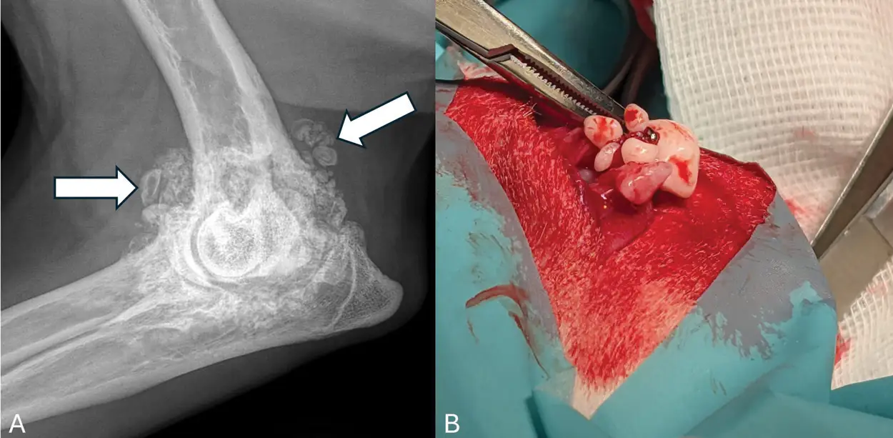
Fig. 2 The left elbow of a 10-year-old, female-neutered Labrador Retriever, 33kg after receiving 18 doses of bedinvetmab (Librela®/Berensa®) for elbow osteoarthritis secondary to elbow dysplasia. Radiograph (A), note the severe atypical new bone formation that extends well beyond the articular margins that has the appearance of severe, coalescing mineralization of the periarticular soft tissues (white arrow). An arthrotomy (B) revealed several intra-articular collections of discrete but coalescing mineralized bodies attached to the synovium by thin fibrous pedicles. Histopathology of the removed tissue revealed irregular nodules which consist of disorganized islands of cartilage and bone. Within the centre of the lesions, there was adipose tissue within the bone marrow spaces. There was also dense fibrous connective tissue surrounding these nodules. A histopathological diagnosis of chondro-osseous metaplasia was made.
To date, no peer-reviewed reports have documented comparable changes in cats following injections of frunevetmab. However, the authors are aware of several feline cases exhibiting similar radiographic features.
Importantly, evidence from human trials and veterinary reports indicates that RPOA can develop in joints that were radiographically unremarkable at treatment initiation. For example, bedinvetmab was prescribed for elbow osteoarthritis, yet catastrophic structural failure subsequently occurred in both tarsal joints– sites not previously identified as clinically affected. These observations suggest that NGF inhibition may reduce the threshold for structural failure in joints undergoing active, even subclinical, degenerative processes.
Although not formally defined, the U.S. Food and Drug Administration’s characterization of RPOA as ‘degenerative joint pathology falling well outside the natural history of osteoarthritis’ is applicable to small animals (Supplementary Material, available in the online version only). However, given the radiographic differences between human RPOA and reported veterinary findings, an alternative terminology– such as ‘aNGFmab-induced rapidly progressive osteoarthritis’–could be considered. Formal definition and validation of such nomenclature should be addressed in future investigations.
An important role of NGF has been shown in cartilage and bone homeostasis as well as soft tissue healing via the tropomyosin receptor kinase A signalling pathway. It has also been shown to mediate the formation of heterotopic mineralization following trauma, and blocking NGF has been discussed as a therapeutic target to prevent such developments.
However, the role of NGF and its signalling pathways is complex, and other studies have shown that perturbation of NGF signalling, using both an NGF signalling agonist and an inhibitor, resulted in a strong induction of chondrocyte calcification, indicating that proper nerve growth factor-tropomyosin receptor kinase A signalling is important for articular cartilage homeostasis. Further study is, therefore, required to determine the exact aetiopathophysiology of aNGFmab-induced rapidly progressive osteoarthritis and help identify risk factors to enable better case selection for treatment and reduce the incidence of ADE.
While aNGFmabs were not approved for human use based on the work by 250 experts assessing 39 trials involving approximately 18,000 patients and 50,000 radiographs, such large-scale, prospective, long-term premarketing structural safety studies in osteoarthritic populations of dogs and cats are not required in veterinary medicine. Therefore, ongoing post-marketing monitoring for possible ADE secondary to bedinvetmab and frunevetmab currently rests largely with the point-of-care clinician. Although serial radiography would be usable, it is relatively insensitive and reflects late-stage change, correlating poorly with actual cartilage loss. Given the logistical and financial constraints of repeated imaging, thorough clinical history and physical examination, therefore, remain the principal safeguards for identifying subtle instability or ‘off-target’ lameness that radiographic changes may not reveal until structural damage is advanced. In addition, should a possible initial effect, wean and ‘lack of efficacy’–one of the most common ADE associated with administration of bedinvetmab– be noted, discontinuation of the aNGFmab and thorough assessment of the patient, including radiographic assessment of any joint that appears abnormal upon examination, are heavily recommended.
Continued vigilance and systematic reporting of suspected ADE associated with bedinvetmab and frunevetmab by using readily available documents are essential to accurately characterize risk and safeguard patient welfare. This information will provide valuable guidance to veterinarians, supporting the safe and evidence-based use of these therapies in dogs and cats with osteoarthritis.
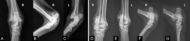
Fig. 3. Radiographic appearance of rapidly progressive osteoarthritic changes in a cat (R¼right; L¼left). This 13-year-old, male-neutered, 6kg Domestic Shorthair cat was treated with two injections of frunevetmab (Solensia®). Initial radiographs (A–C) and radiographs 2 months later (D, E): Right tarsus: Severe periosteal reaction throughout the tarsal joint with severe erosion of subchondral bone surfaces and apparent instability/subluxation of the joint. Left tarsus: Severe periosteal reaction throughout the tarsal joint with severe erosion of subchondral bone surfaces and apparent instability/subluxation of the joint. This has progressed significantly from initial radiographs (A–C). Joint fluid cytology, culture, haematology, blood chemistry did not reveal concerning abnormalities and helped to rule out other aetiologies, including immunemediated arthritis. Histopathology of the synovium showed chronic inflammation and synovial hyperplasia, similar to human histopathological results of rapidly progressive osteoarthritiscases.Thus, frunevetmab-related increased deterioration was established as cause for severe and more rapid progressive changes than would be expected for typical feline osteoarthritis.
Important advances toward this objective have recently been reported, including the presentation of additional data at the Veterinary Orthopedic Society Annual Meeting in March 2026. The authors described 38 cases exhibiting a consistent pattern of ADE in dogs treated with bedinvetmab, with a mean time to onset of 6.5 months. With the exception of a single case, all dogs received the drug at recommended dosages, frequently in combination with non-steroidal anti-inflammatory drugs, and developed a subacute, progressive and destabilizing arthropathy characterized by imaging findings including joint laxity, osteophytosis, effusion and subchondral alterations. Clinical outcomes were frequently severe, with a substantial proportion of affected dogs requiring surgical intervention, and some progressing to euthanasia or limb amputation due to compromised quality of life, underscoring the potential clinical relevance of these observations.
Click here to view the full publication
Author Affiliations:
- Small Animal Surgery Locum, PLLC, Dallas, Texas, United States.
- Spry Companions Veterinary Orthopedic Specialists, Denver, Colorado, United States.
- Michigan State University, Department of Small Animal Clinical Sciences, College of Veterinary Medicine, East Lansing, Michigan, United States.
- Greenside Veterinary Practice, Greenside Referrals, Melrose, Roxburghshire, United Kingdom.
- Department of Molecular Biomedical Sciences, North Carolina State University, Raleigh, North Carolina, United States.